Showing posts with label GIT. Show all posts

Wednesday, February 17, 2010

Ostomy Covers and Bags (Ileostomy, Colostomy): I Bet You Didn't Think Of This


________________________________
Ostomy covers are just one in a long series of supplies necessary in the care of colostomy and ileostomy patients. The words ileostomy and colostomy refer to the procedure ending up with a bag coming out of the abdomen that fills with stool. They are usually placed in folks who have lost the distal portion of their colon (colostomy) or when the entire colon has been removed or when the small bowel must be diverted (ileostomy). They can sometimes be reversed (called a take down) when the underlying reasons for placing them are fixed (such as massive decubiti ulcers).

All ostomy sites, whether they are ileostomy or colostomy sites, must be protected to ensure their survival. Ostomies require care to prevent ulcers at the exit site. They can get ischemic and and swollen and obstructed. Sometimes they need to be surgically revised. They need to be kept clean at the surface to prevent infections. They should be kept dry while in the shower or bath.

And that stuff costs money. You can find ostomy covers and ostomy bags in all assorted sizes and colors at online stores or at your local medical supply company. In fact you can even find designer ostomy covers and bags. Maybe this represents a growth opportunity for the higher end designer bag makers Coach, Kate Spade and Loui Vitton.

Designer ostomy covers and bags could take future growth projections for these higher end bag makers to a whole new level. The growth opportunities are enormous for an aging and wealthy population who cares less about the bag on their shoulder and more about the bag on their belly.

But don't forget about the little ones out there. Perhaps even Disney could market Minnie ostomy covers and bags for the little kids out there stricken with illness. You could have the best first grade show and tell ever with your Goofy ostomy cover. Most kids bring a trinket . Your kid shows his Goofy ostomy cover.

Think of the other possibilities. Hospitals could throw designer ostomy cover and bag parties as a new way to fund their daily operations as the Medicare National Bank goes belly up. Or perhaps, in addition to pet therapy dogs, an ala carte menu of optional services for patients could include offering them special ostomy covers for their ostomy bags.

Look at the spirit of cooperation between hospitals and hospitalists and you can understand the power of market economics in medicine. Many doctors are troubled by today's EMRs. They don't know it yet, but someday EMRs will be their saving grace. And it has nothing to do with making medical care better or more efficient. In fact, as insurance cheapens the value of medical education, EMRs will become nothing more than a tool for direct marketing.

Medical doctors could go the way of chiropractic marketing and show up at trade shows and State Fairs peddling a whole array of ostomy covers and ostomy bags to a public willing to spend their money on everything but actual health care. Or better yet primary care doctors, internists and gastroenterologists could give the knock off jewelery and purse parties a run for their money. These doctors have a focused clientele right at their finger tips. If they have a patient panel and an EMR, they have a whole new and exciting business opportunity just waiting for them.

They just have to run an EMR comparison for those patients with an ostomy and send them an invitation to their private party selling designer or knockoff designer ostomy covers and bags. You think Pampered Chef parties are out of control. Wait until you have a million doctors inviting you to their ostomy cover parties. Just you wait. It's going to happen.

I don't think anyone in Washington thought of the unintended consequences of a declining Medicare payment model coupled with the explosion of EMR technology. Doctors are business owners with an entrepreneurial spirit. They will always search for alternative sources of income as insurance decimates their bottom line. Third party insurance companies just don't want to pay for health care anymore. I witnessed that first hand with my physician review experience. And selling ostomy covers is just one way for doctors to survive. But it won't stop with colostomy covers and ileostomy bags.

Someday you'll have pulmonologists using their EMR for direct marketing of their designer tracheostomy parties. You'll have vascular surgeons using their EMR for direct marketing of their designer stump parties. You'll have opthalmologists taking up designer eyeball tattoos with the help of their EMR. And you'll have primary care doctors doing botox. Oh wait, they are already doing that.

That's the future of the EMR. It has nothing to do with medical care. In fact, the future driving revenue for doctors won't be medical care at all. Insurance killed that idea. It will be medical supplies. Instead of using the EMR to streamline health care, doctors will use it to data mine their patients for the direct to consumer advertising and marketing of goods and services. You heard it here first. You thought the drug companies were bad. Just wait until doctors figure out the power of the EMR.

The whole idea of doctors using their office and patient panel as a direct marketing force de jour lies on the premise that patients are more willing to pay for things than for service. And that's true for most of them. You see them pulling up in their full sized SUVs with the shiny 26 inch rims, their smart phones, their $200 jeans and their cigarettes sticking out of their designer purse. You know they're willing to pay for image. But they won't pay a $10 copay to evaluate their diabetes, heart failure, COPD, atrial fibrillation, high cholesterol, leg pains, dizziness, shakes, dry skin, diarrhea, gas, rash and sore throat all in one visit. You want to survive the future of health care? I suggest you get yourself an EMR and start selling something tangible.
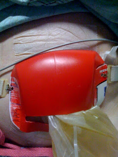
Ostomy-Colostomy-Ileostomy-Designer-Bag
And for the patients who don't want anything to do with designer ostomy covers? What do we do about them? How do we make money on them in the future? We don't. But we take great pleasure in discovering the lengths people go to save a buck. And we enjoy the creative nature of the human mind. Like this awesome guy who made his own designer ostomy cover (actually for a urostomy) by cutting out the back side of a bottle of liquid laundry detergent. I suppose you could say his ostomy will never get dirty. Perhaps this guy should start his own business selling ostomy covers to poor people, so he can then buy one from you at your exclusive invitation only ostomy covers party.

Now there's a thought...
Blog Widget by LinkWithin

Tuesday, February 16, 2010

Prostate problem probed

Pinpointing prostate problems The chemical cousin of magnetic resonance imaging, MR spectroscopy, could be used to pinpoint the exact location of prostate cancers and to determine the aggressiveness of a tumour without major surgical intervention, according to research published in the journal Science Translational Medicine.

“Magnetic resonance (MR) spectroscopy which can analyse the biochemistry rather than the physical structure of tissues could give oncologists a better way to home in on prostate cancers at the early stages of growth and so ultimately improve treatment success rates,” team leader Leo Cheng of Harvard U told me.

Twenty-year old HIV problem – Science journalists (and press officers alike) are often lambasted for using the word breakthrough, but when twenty years of research culminates in a development with the potential to change the way drug research for HIV/AIDS is undertaken, we can be forgiven, surely. An X-ray diffraction study of the enzyme integrase has led to a breakthrough in our understanding of how retroviruses replicate. The structural results lay bare a problem that scientists have been trying to solve for more than two decades.

Sniffing out the Tempranillo – It’s unclear how common fraud is in the wine industry, but certainly there is the opportunity for unscrupulous producers to blend wine of the same variety from different areas and claim it comes from the most well-renowned regions. Now, Australian scientists have developed a new approach to testing the origin of wines based on a sophisticated statistical analysis of the wine’s spectra.

Atomic biodiesel assessment – Metal contaminants in your car’s biofuel will cause the build up of sludge inside the engine and potentially cause it to fail, replacing a burned out engine is not the green option for those hoping to save the environment by burning crops instead of oil in their vehicles. Now, Brazilian scientists have turned to Flame atomic absorption spectrometry (FAAS) to quickly and cheaply determine the metal content of biodiesel with a view to improving quality control on this renewable fuel.

Research Blogging IconWu, C., Jordan, K., Ratai, E., Sheng, J., Adkins, C., DeFeo, E., Jenkins, B., Ying, L., McDougal, W., & Cheng, L. (2010). Metabolomic Imaging for Human Prostate Cancer Detection Science Translational Medicine, 2 (16), 16-16 DOI: 10.1126/scitranslmed.3000513

Everybody knows that colonoscopy is the best test to screen for colorectal cancer and that colonoscopies save lives. Everybody may be wrong. Colonosco

Everybody knows that colonoscopy is the best test to screen for colorectal cancer and that colonoscopies save lives. Everybody may be wrong. Colonoscopy is increasingly viewed as the gold standard for colorectal cancer screening, but its reputation is not based on solid evidence. In reality, it is not yet known for certain whether colonoscopy can help reduce the number of deaths from colorectal cancer. Screening with fecal occult blood testing (FOBT) and flexible sigmoidoscopy are supported by better evidence, but questions remain. It seems our zeal for screening tests has outstripped the evidence.

Statistics show that the life-time risk for an adult American to develop colorectal cancer (CRC) is approximately 6%. Colorectal cancer is the second leading cause of cancer deaths in the United States. In the US there are currently 146,970 new cases and 50,630 deaths each year. Between 1973 and 1995, mortality from CRC declined by 20.5%, and incidence declined by 7.4% in the United States.

The US Preventive Services Task Force (USPSTF) recommends screening for colorectal cancer (CRC) using fecal occult blood testing, sigmoidoscopy, or colonoscopy, in adults, beginning at age 50 years and continuing until age 75 years.

The American Cancer Society divides the available tests into these two categories and makes these recommendations for frequency of testing:

Tests that find polyps and cancer

  • flexible sigmoidoscopy every 5 years*
  • colonoscopy every 10 years
  • double contrast barium enema every 5 years*
  • CT colonography (virtual colonoscopy) every 5 years*

Tests that mainly find cancer

  • fecal occult blood test (FOBT) every year*,**
  • fecal immunochemical test (FIT) every year*,**
  • stool DNA test (sDNA), interval uncertain*

Screening by colonoscopy seems to make more sense than other screening methods, because you can actually see the entire inside of the colon. Colon cancer is preceded by polyps and adenomas that progress to cancer. When a polyp is seen, it can be removed during the procedure. In this study, colonoscopic polypectomy resulted in a lower than expected incidence of colorectal cancer. But other studies suggest that the progression to cancer is not a steady process, and that adenomas may regress.

There is good evidence here and here that any benefit of colonoscopy is restricted to left-sided colon cancers, with no impact on right-sided colon cancer; we don’t understand why. Some possible explanations are discussed here.

There are pros and cons to each of the different screening tests. Barium enemas and CT virtual colonoscopy involve significant doses of radiation. Colonoscopy only needs to be done every 10 years, but it involves an uncomfortable bowel prep, requires sedation, can cause serious complications like bowel perforation, and is unacceptable to some patients. FOBT screening is painless and harmless but has a lot of false positives and requires annual testing. Getting patients to come back every year for FOBT is problematic. Compliance and cost must be considered. Colonoscopy is expensive and there are not enough colonoscopists to screen everyone.

Apart from all those peripheral considerations, what do we know about the bottom line: the ability of each screening method to prevent deaths from colon cancer? According to the National Cancer Institute,

  • Studies have shown that FOBT, when performed every 1 to 2 years in people ages 50 to 80, can help reduce the number of deaths due to colorectal cancer by 15 to 33 percent.
  • Studies suggest that regular screening with sigmoidoscopy after age 50 can help reduce the number of deaths from colorectal cancer, perhaps by as much as 50%, but the quality of evidence is not as good as for FOBT.
  • it is not yet known for certain whether colonoscopy can help reduce the number of deaths from colorectal cancer.

No randomized controlled trials have tested whether colonoscopy reduces the incidence of CRC. Support for the role of colonoscopy in CRC prevention derives from indirect evidence and observational studies.

There is an excellent review of all the pertinent studies here. Even though studies show that screening can reduce disease-specific mortality from colorectal cancer, there is little evidence that it reduces all-cause mortality. So as far as we know, screening probably won’t prolong your life. It seems like it should: I don’t understand why it doesn’t, and it bothers me. This certainly isn’t the message we’re getting from the media and from the medical profession.

I’m guessing that if the appropriate studies were done and the technique of colonoscopy were optimized, a reduction in colon cancer deaths would be demonstrated. I’m guessing that colonoscopy would detect more cancers and precancerous lesions than FOBT or sigmoidoscopy, but I’m wondering whether the benefits of colonoscopy would outweigh the additional cost and risks compared to other screening methods. And I’m disturbed that a reduction in all-cause mortality has not yet been clearly shown for any screening method. We need more research to help us understand these issues.

Pending better evidence, I support the current USPSTF recommendations. I think patients should be told the pros and cons and choose which screening test they prefer. I have chosen annual FOBT for

Thursday, February 11, 2010

Midgut pain due to an intussuscepting terminal ileal lipoma: a case report

Journal of Medical Case Reports 2010, 4:51 doi:10.1186/1752-1947-4-51
Noormuhammad O Abbasakoor (abbasakn@tcd.ie)
Dara O Kavanagh (dara_kav@hotmail.com)
Diarmaid C Moran (morandiarmaid@gmail.com)
Barbara Ryan (abbasakn@tcd.ie)
Paul C Neary (paulcneary@msn.com)
ISSN 1752-1947
Article type Case report
Submission date 19 September 2009
Acceptance date 11 February 2010
Publication date 11 February 2010
Article URL http://www.jmedicalcasereports.com/content/4/1/51
This peer-reviewed article was published immediately upon acceptance. It can be downloaded,
printed and distributed freely for any purposes (see copyright notice below).
Articles in JMCR are listed in PubMed and archived at PubMed Central.
For information about publishing your research in JMCR or any BioMed Central journal, go to
http://www.jmedicalcasereports.com/info/instructions/
For information about other BioMed Central publications go to
http://www.biomedcentral.com/
Journal of Medical Case
Reports
© 2010 Abbasakoor et al. , licensee BioMed Central Ltd.
This is an open access article distributed under the terms of the Creative Commons Attribution License (http://creativecommons.org/licenses/by/2.0),
which permits unrestricted use, distribution, and reproduction in any medium, provided the original work is properly cited.Midgut pain due to an intussuscepting terminal ileal lipoma: a case report

Noormuhammad O Abbasakoor
1
, Dara O Kavanagh1
, Diarmaid C Moran1
, Barbara Ryan2
,
Paul C Neary1*


Addresses:
1
Division of Colorectal Surgery, Adelaide and Meath Incorporating the
National Children’s Hospital, Tallaght, Dublin 24, Ireland.
2
Department of
Gastroenterology, Adelaide and Meath Incorporating the National Children’s Hospital,
Tallaght, Dublin 24, Ireland.

*Corresponding author

PCN: paulcneary@msn.com Abstract
Introduction
The occurrence of intussusception in adults is rare. The condition is found in 1 in 1300
abdominal operations and 1 in 100 patients operated for intestinal obstruction. The child
to adult ratio is 20:1.

Case presentation
A 52-year-old Irish Caucasian woman was investigated for a 3-month history of
intermittent episodes of colicky midgut pain and associated constipation. Ileocolonoscopy
revealed a pedunculated lesion in the terminal ileum prolapsing into the caecum.
Computed tomography confirmed a smooth-walled, nonobstructing, low density
intramural lesion in the terminal ileum with secondary intussusception. A laparoscopic
small bowel resection was performed. Histology revealed a large pedunculated polypoidal
mass measuring 4x2.5x2cm consistent with a submucosal lipoma. She had complete
resolution of her symptoms and remained well at 12-month follow-up.

Conclusion
This case highlights an unusual cause of incomplete small bowel obstruction successfully
treated through interdisciplinary cooperation. Ileal lipomas are not typically amenable to
endoscopic removal and require resection. This can be successfully achieved via a
laparoscopic approach with early restoration of premorbid functioning.


Introduction
Neoplasms of the small intestines are rare [1]. Gastrointestinal lipomas are benign
tumors that can occur in the small bowel but occur most commonly in the colon. The
majority are asymptomatic and are detected incidentally on abdominal imaging. Removal
is warranted if tissue diagnosis is deemed essential or if severe symptomatology, such as
pain or bleeding, exists [2].

We report a case of terminal ileal lipoma causing intermittent intussusception in a 52-
year-old woman. The lipoma was diagnosed at ileocolonoscopy and successfully removed
through laparoscopy. A review of the literature on small bowel intussception and
gastrointestinal (GI) lipomas is also presented in this report.

Case presentation

A 52-year-old Irish Caucasian woman presented with a three-month history of
intermittent central abdominal pain and constipation. She did not describe
gastrointestinal bleeding or weight loss. She previously underwent a transabdominal
hysterectomy for menorrhagia. Her physical examination was unremarkable. Initial
investigations, such as blood tests, abdomen ultrasound and gastroscopy were
unremarkable. Ileocolonoscopy revealed a pedunculated terminal ileal lesion prolapsing
into her caecum. Computed tomography (CT) of her abdomen and pelvis demonstrated a
smooth-walled, low-density, intramural lesion in the terminal ileum. It measured
3.2x1.6cm. The ileum at the proximal end of the lesion was mildly dilated with a
centrally placed narrowed channel of contrast, which was consistent with an
intussusception possibly secondary to an intramural lipoma. There was no evidence of
obstruction (Figure 1).

She underwent an elective laparoscopic small bowel resection and stapled functional
end-to-end anastomoses. On macroscopy the lesion appeared as a large pedunculated
polypoid mass measuring 4x2.5x2cm with focal mucosal ulceration (Figure 2). Microscopy revealed a submucosal lipoma with blunting of the overlying mucosal villi and
pyloric gland metaplasia. She made an uneventful recovery and was discharged home on
the fourth postoperative day. She returned to work on the 12th
postoperative day. She
remained free of symptoms at three-month follow-up.

Discussion

Lipomas are benign tumors of mesenchymal origin. They are the second most common
benign tumors in the small intestine and account for 10% of all benign gastrointestinal
tumors and 5% of all gastrointestinal tumors. They are predominantly submucosal and
protrude into the lumen [2]. Occasionally, they arise in the serosa. Gastrointestinal
lipomas are most commonly located in the colon (65% to 75%, especially on the right
side), small bowel (20% to 25%), and occasionally in the foregut (<5%) [2]. Lipomas
are largely asymptomatic. Major presenting features are intestinal obstruction and
hemorrhage [3].

Intussusception in adults is a rare entity that it is generally caused by definable
intraluminal pathology [4]. Diagnosis can be challenging. Intussusception is classified
according to its gastrointestinal location: enteric, ileocaecal, or colonic [4]. In ileocaecal
intussusceptions, the ileocaecal valve acts as the lead point. The ileum
(‘intussusceptum’) telescopes into the colon (‘intussuscipiens’) through the ileocaecal
valve [5, 6]. Intussusception leads to the development of venous and lymphatic
congestion, which results in intestinal edema. If not treated promptly, the arterial blood
supply to the bowel will be compromised, thus leading to ischaemia, perforation and
peritonitis [4]. Only 5% of all intussusceptions occur in adults [7]. In 90% of these cases
a predisposing lesion is identified [7]. This is contrary to intussusception in the pediatric
population where an organic lesion is found in only 10% of documented cases [3]. In
adults, it is important to differentiate between small bowel and colonic intussusception.
In 63% of cases of small bowel intussusceptions, a benign underlying lesion can be found. Meanwhile, a malignant etiology has to be expected in 58% of cases of large
bowel intussusceptions [8].

Lipomas can be diagnosed through conventional endoscopy, capsule endoscopy, barium
studies and, most importantly, CT. Typical endoscopic features are smooth, yellowish
surface with pedunculated or sessile base, as seen in this case. Other endoscopic
characteristics are the ''cushion sign'' and ''naked fat sign” [2]. CT usually reveals a
smooth, well-demarcated sausage-shaped mass. It may also reveal associated
intussusception if present [5]. Capsule endoscopy and digital balloon endoscopy are
newer means for diagnosing lipomas and are particularly helpful in cases involving small
bowel lipomas [2]. Associated intussusception can be confirmed on contrast enema
(‘crescent sign’), CT and magnetic resonance imaging (MRI). Multislice CT facilitates the
assessment of vascular supply to the affected bowel loop in cases of intussusception
where impending ischemia is suspected [4].

The treatment for lipomas depends on the clinical manifestations. Indications for their
removal include intestinal obstruction, hemorrhage and malignant potential [4]. There is
a theoretical risk of sarcomatous change but this has rarely been documented in the
literature [1]. Endoscopic removal is possible but potentially complicated. In view of the
submucosal location, there is an inherent risk of perforation [9]. Furthermore, lipomas
have high water content, which means a large amount of cautery is necessary to achieve
effective hemostasis [9]. Surgery can be performed through laparoscopy or via an open
approach. The type of resection and anastomosis depends on the location, bowel wall
integrity, and vascular supply of the lipoma [6]. Elective laparoscopic resection of
lipomas is the treatment of choice with the concomitant benefits of laparoscopic surgery,
such as shorter duration of hospital stay, less postoperative pain, early restoration of
(GI) function and good cosmesis [6].

Conclusion In this case, we illustrate the importance of a thorough interdisciplinary evaluation of
patients with midgut abdominal pain. It highlights the diagnostic values of CT scanning
and completed ileocolonoscopy. Despite preoperative localization, laparoscopy facilitates
a thorough evaluation of the intraperitoneal contents and therapeutic resection of the
affected segment. This report confirms the recognized benefits of laparoscopic surgery
with associated early return to premorbid functioning. In patients with persistent
episodes of incomplete intestinal obstruction, atypical causes, such as the etiology we
describe here, should be considered.

Consent
Written informed consent was obtained from our patient for publication of this case
report and any accompanying images.

Competing interests
The authors declare they have no competing interests.

Authors’ contributions
NOA contributed in collecting the requisite literature and wrote the case report. DOK also
collected the requisite literature and reviewed the literature. DCM also contributed in
collecting the requisite literature. BR and PCN were involved in the diagnosis of our
patient. PCN also performed the surgery. All authors read and approved the final
manuscript. References
1. Rathore MA, Andrabi SI, Mansha M: Adult intussusception: a surgical
dilemma. J Ayub Med Coll Abbottabad 2006 Jul-Sep, 18(3):3-6.
2. Chou JW, Feng CL, Lai HC, Tsai CC, Chen SH, Hsu CH, Cheng KS, Peng CY, Chung
PK: Obscure gastrointestinal bleeding caused by small bowel lipoma.
Inter Med 2008, 47:1601-1603.
3. Balik AA, Ozturk G, Aydinli B, Alper F, Gumus H, Yildirgan MI, Basoglu M:
Intussusception in adults. Acta Chir Belg 2006 Jul-Aug, 106(4):409-412.
4. Lin HH, Chan DC, Yu CY, Chao YC, Hsieh TY: Is this a lipoma? Am J Med 2008
Jan, 121(1):21-23.
5. Michael A, Dourakis S, Papanikolaou I: Ileocaecal intussusception in an adult
caused by a lipoma of the terminal ileum. Ann Gastroenterol 2001,
14(1):56-59.
6. Takaaki T, Matsui N, Hiroshi K, Takemoto Y, Oka K, Seyama A, Morita T:
Laparoscopic resection of an ileal lipoma: report of a case. Surg Today
2006, 36:1007-1011.
7. Meshikhes AW, Al-Momen SA, Al Talaq FT, Al-Jaroof AH: Adult intussusception
caused by a lipoma in the small bowel: report of a case. Surg Today 2005,
35(2):161-165.
8. Oyen TL, Wolthuis AM, Tollens T, Aelvoet C, Vanrijkel JP: Ileo-ileal
intussusception secondary to a lipoma: a literature review. Acta Chir Belg
2007, 107:60-63.
9. Yoshimura H, Murata K, Takase K, Nakano T, Tameda Y. A case of lipoma of the
terminal ileum treated by endoscopic removal. Gastrointestinal
Endosc.1997 Nov, 46(5):461-463.

Figure legends
Figure 1. Contrast-enhanced computed tomography scan of the abdomen demonstrates
a smooth-walled, low-density intramural lesion. It measures 3.2x1.6cm. The ileum at the proximal end of the lesion is mildly dilated with a centrally placed narrowed channel of
contrast consistent with an intussusception.

Figure 2. Macroscopic view of a large pedunculated polypoid mass arising from the
luminal surface of the ileal resection specimen. Appearances are consistent with a
lipoma.

Figure 1

Wednesday, February 10, 2010

Baby broccoli 'controls gut bug'


Helicobacter pylori
The bacteria lives in the gastrointestinal tract

Eating a daily portion of broccoli sprouts could help tame the H. pylori bacteria, linked to stomach ulcers and even cancer, research suggests.

The study in Cancer Prevention Research of 50 people in Japan found eating 2.5 ounces of broccoli sprouts each day for two months may confer some protection.

They contain sulforaphane, previously found to act as an antibiotic.

UK experts said while sprouts may have an effect on the bug, they were likely to make "no difference" to cancer risk.

This small study shows that eating broccoli sprouts might reduce levels of H. pylori infection
Nell Barrie, Cancer Research UK

In the study, an international team of scientists gave half the group a daily portion of broccoli sprouts and the rest alfalfa sprouts, which do not contain sulforaphane.

In those who ate broccoli sprouts, levels of a marker of H. pylori in human stools called HpSA was cut by over 40%.

There was no HpSA level change in those who ate alfalfa sprouts.

In people who ate broccoli sprouts, HpSA levels had returned to pre-treatment levels eight weeks after people stopped eating them.

The researchers say this suggests that although the sprouts can dampen down H. pylori, they do not eradicate it.

Sprout smoothies

Dr Jed Fahey, of Johns Hopkins University in the US who led the study, said: "The fact that the levels of infection and inflammation were reduced suggests the likelihood of getting gastritis and ulcers and cancer is probably reduced."

It was Dr Fahey who discovered the sprouts contained sulforaphane early this decade. He is a co-founder of a company licensed by The Johns Hopkins University to produce broccoli sprouts. A portion of the proceeds is used to help support cancer research.

His team also carried out tests on mice infected with H. pylori, giving them broccoli-sprout smoothies for eight weeks.

The number of H. pylori bacteria in the mice's stomachs fell significantly - but did not change in infected mice that only drank plain water.

A second group of H. pylori-infected mice were genetically engineered to lack the Nrf2 gene that activates protective enzymes.

They failed to respond in the same way to the sprout-smoothie diet.

Nell Barrie of Cancer Research UK said: "This small study shows that eating broccoli sprouts might reduce levels of H. pylori infection.

"We know that H. pylori is a major risk factor for stomach cancer but only three in a 100 people with the infection will develop the disease, so there are clearly other factors at work.

"This means we can't conclude that eating broccoli sprouts makes any real difference to the chance of getting stomach cancer. "

Inflammatory bowel disease hikes blood clots

Study finds people with Crohn’s disease or ulcerative colitis face highest risk during flare-ups

By Nathan Seppa


People with inflammatory bowel disease are at an increased risk of developing blood clots, and this risk is highest during a flare-up, researchers report online February 9 in the Lancet. Although hospitalized IBD patients have been known to have problems with clotting, the new study finds a surprisingly high risk in people dealing with an episode outside the hospital setting.

IBD includes Crohn’s disease and ulcerative colitis. Although medication can bring IBD under control and keep it in remission temporarily, relapses of the disease are common and can debilitate a person. Patients often require steroid medication to knock back inflammation that causes the abdominal pain, diarrhea, vomiting and blood in the stool that mark the condition.

In the new study, Matthew Grainge and his colleagues at the University of Nottingham in England analyzed medical records dating from 1987 to 2001 of more than 13,000 IBD patients and a control group of 71,000 people who didn’t have IBD. People in both groups were in their mid-40s on average and had comparable weights and smoking histories, factors that also influence blood clot formation. The researchers excluded people whose records showed a prescription for blood thinners or who might have taken steroids for other ailments, such as asthma.

The scientists found that hospitalized IBD patients fending off a flare-up were three times more likely to develop venous thrombosis — a blood clot in a vein — than were hospitalized people in the control group without IBD, a finding that matches past reports. But IBD patients dealing with a flare-up outside a hospital setting were 16 times more likely than the nonhospitalized control group to develop a venous thrombosis. In absolute terms, people with IBD in the hospital were still at greater risk than those who were treated as outpatients.

Even while in remission, IBD patients were twice as likely as the people who didn’t have the disease to develop such a clot.

A venous thrombosis that occurs in a large vein in a leg or arm, a condition called deep-vein thrombosis, can cause pain and swelling. The chief risk is that the clot will dislodge and cause a pulmonary embolism, a blockage of one of the main arteries in the lung. That can lead to chest pain, breathing problems, loss of oxygen, abnormal heart rate, sinking blood pressure, collapse and even death. On rare occasions, according to case studies, these clots can lodge in the brain and cause strokes.

The reason for increased clot risk in IBD patients remains a mystery. Steroid use might contribute to it, the authors say, but additional risk has not shown up in other groups using steroids, including people with asthma or rheumatoid arthritis.

Some scientists hypothesize that the systemic inflammation that characterizes IBD might trigger clotting, but it’s not clear how, says Geoffrey Nguyen, a gastroenterologist at the University of Toronto.

People in hospitals tend to be sicker and therefore have a greater risk of clotting whether they have IBD or not, Nguyen says. This may account in part for why the hospitalized IBD patients were only three times more likely to have clots than their hospitalized counterparts while the risk for the nonhospitalized IBD patients was much higher compared to nonhospitalized controls. Also, hospitalized IBD patients in this study might have received short-term regimens of blood thinners that didn’t show up on the prescription records analyzed, he notes.

In any case, Nguyen says, this study should help doctors in treating people who have IBD. “It’s important for clinicians to really instruct patients that they have a condition that increases the risk of clots,” he says. To that end, he suggests that the kind of support stockings used to minimize clot formation on long airplane flights are helpful and inexpensive, Nguyen says. And most important, patients need to report promptly any swelling and pain in extremities, as these may indicate venous thrombosis, he says

Colon Detoxification

The internet is full of colon cleansing methods that tout the benefits of colon detox. I saw one website that showed long "worms" that live for years in the colon that "need" to be removed with special expensive potions. One of the most common questions for GI doctors is about colon cleansing and if it is beneficial. I don't know any physicians who believe the colon needs "detoxification" or special cleansing, but until now I didn't have a scientific way to answer that question from patients.

A study from the Am J. Gastroenterology now gives us the answer. The study authors looked at all relevant articles published between 1966 and 2008. They blinded the articles and measured outcomes and adverse events.

No research articles addressed the effect of colon cleansing on general health or on specific conditions such as hypertension, asthma, irritable bowel syndrome, arthritis, sinus congestion or alcoholism, despite the fact that these conditions are mentioned in ads. They did find adverse events such as electrolyte imbalances, septicemia, colitis, rectal perforation and death. One article reported an outbreak of amebiasis (amoeba infection) attributed to colonic irrigation therapy with at least 36 patients infected. Ten required colectomy and six patients died.

There was no good quality published evidence of any health benefit from colonic cleansing and many reports of adverse events. This practice has a large following with many colonic substances including coffee enemas and other herbs administered in the enema fluid.

The colon is designed to absorb nutrients and remove waste. The best way to keep your colon and GI tract healthy is to eat fruit, vegetables and fiber, stay hydrated and active. Special potions or enemas not needed.
Related Posts with Thumbnails